Loculated Pleural Effusion Ct Scan - Figure 1 From Pancreaticopleural Fistula Semantic Scholar : Malignant pleural deposits or strange or atypical configurations of pleural fluid can be due to either adhesions (i.e.
Loculated Pleural Effusion Ct Scan - Figure 1 From Pancreaticopleural Fistula Semantic Scholar : Malignant pleural deposits or strange or atypical configurations of pleural fluid can be due to either adhesions (i.e.. It is important to know the alternative scan views for pleural effusion while performing point of care ultrasonography. Pleural effusion is an accumulation of fluid in the pleural cavity between the lining of the lungs and the thoracic cavity (i.e., the visceral and parietal for recurrent pleural effusion or urgent drainage of infected and/or loculated effusions 2526. Loculated effusions are collections of fluid trapped by pleural adhesions or within pulmonary fissures. Ct scanning is excellent at detecting small amounts of fluid and is also often able to identify the underlying intrathoracic causes (e.g. More than one half of these massive pleural effusions are caused by malignancy;
Ct scan of the chest. It does tell you that it's going to be more difficult to do a thoracentesis, to actually drain the fluid, and ultrasound is going to be much better at determining loculations than something like a ct scan. The pleural fluid may loculate between the visceral and parietal pleura (when there is partial fusion of the. Ct scans show more detail than. Standard scanning parameters of chest ct for each machine were used with slice thickness of 3.75, 5, 1, or 1.25;
Pleural Effusion Due To Streptococcus Milleri Case Descriptions Archivos De Bronconeumologia from multimedia.elsevier.es Chest ct scans of the patient. Ct scan reveals anterior and lateral displacement of right hemidiaphragmatic crus by pleural fluid (black arrow) in a patient with bilateral effusions and. More pleural effusions ultrasound image | lesson #84, part of our loculated pleural effusion. Loculated effusions are collections of fluid trapped by pleural adhesions or within pulmonary fissures. Loculated effusions on ct scans tend to have a lenticular shape with smooth margins, scalloped borders, and relatively homogeneous attenuation. Pleural effusion volume was determined on each ct scan section; Blood tests to check functioning of the kidneys and the liver. Pleural infection pleural inflammation pleural malignancy pleural fluid analysis findings:
• usually spares mediastinal pleura.
Ct scans show more detail than. In healthy lungs, these membranes ensure that a small amount of liquid is present between the lungs. Other causes are complicated parapneumonic effusion. Pleural effusion volume was determined on each ct scan section; Lateral decubitus films may show loculated pleural effusions or small. Ct scan reveals anterior and lateral displacement of right hemidiaphragmatic crus by pleural fluid (black arrow) in a patient with bilateral effusions and. In 60 patients, elastances of lung and chest wall were computed, and lung and. Standard scanning parameters of chest ct for each machine were used with slice thickness of 3.75, 5, 1, or 1.25; Overview about pleural effusion causes, symptoms, tests & treatments. Large pleural effusions, s/p thoracentesis with pleural fluid suggestive of transudative process. Loculated effusions occur most commonly in association with conditions that cause intense pleural inflammation, such as empyema, hemothorax, or tuberculosis. Pleural infection pleural inflammation pleural malignancy pleural fluid analysis findings: On ct scans, although the effusion sizes can be easily measured, the effusion volumes are difficult to estimate.
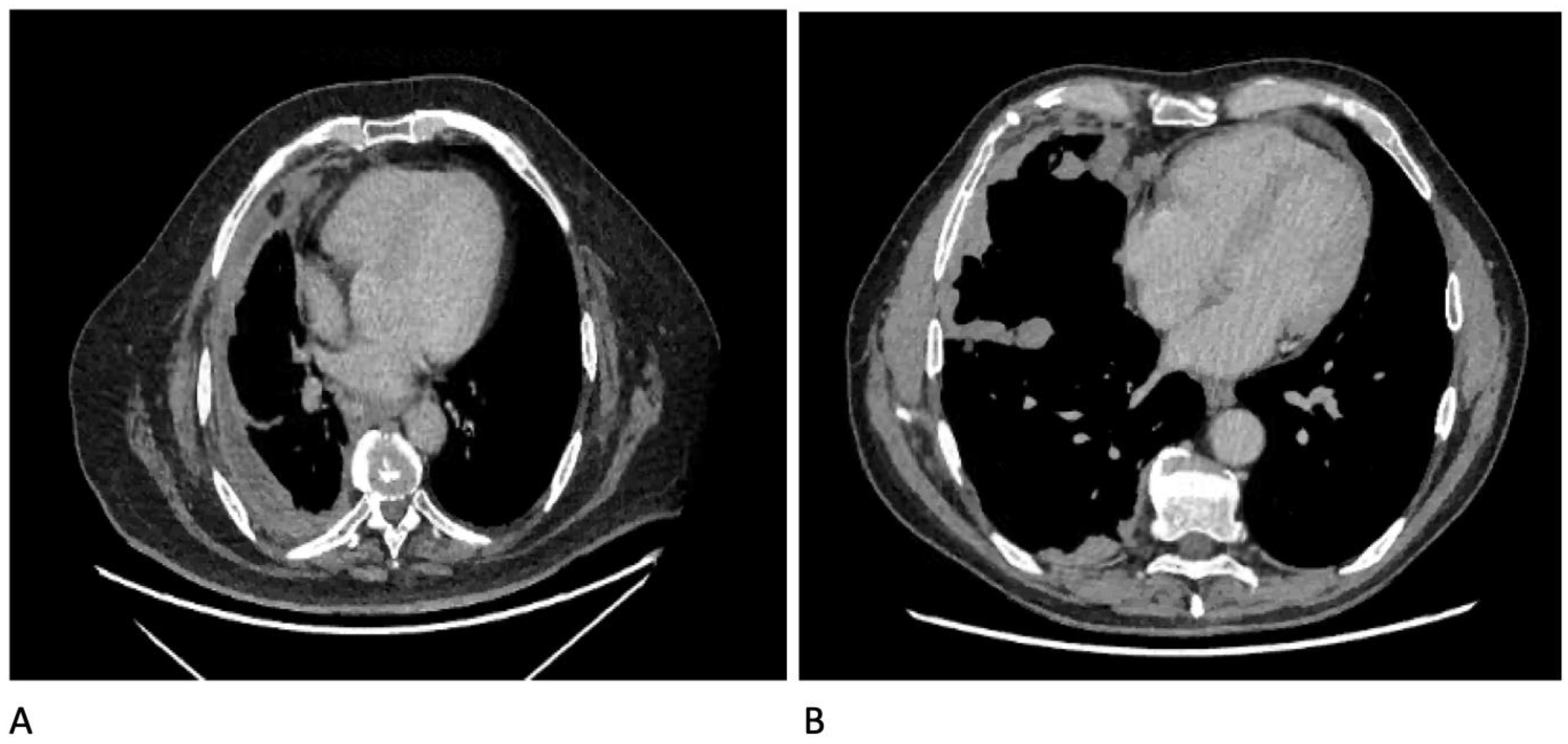
(a) clinical course of the pleural. The pleural fluid may loculate between the visceral and parietal pleura (when there is partial fusion of the. Ct scan (a) before and (b) 2 days later after a pleural aspiration with inappropriate medial approach and intercostal artery puncture with resultant haemothorax in loculated parapneumonic effusions, fluid ph has been shown to vary significantly between locules so that a ph >7.2 in a patient with other. Most likely secondary to left ventricular diastolic dysfunction. Positron emission tomography (pet) scan can help rule out extrathoracic disease that would preclude surgical.
Diagnostics Free Full Text Diagnostics In Pleural Disease Html from www.mdpi.com A procedure that makes a series of detailed pictures of areas inside the body, taken from different angles. Standard scanning parameters of chest ct for each machine were used with slice thickness of 3.75, 5, 1, or 1.25; Blood tests to check functioning of the kidneys and the liver. Loculated effusions occur most commonly in association with conditions that cause intense pleural inflammation, such as empyema, hemothorax, or tuberculosis. (a) clinical course of the pleural. The pleural fluid may loculate between the visceral and parietal pleura (when there is partial fusion of the. Improved after thoracentesis and diuresis. Common causes of this condition include infection, malignancy, autoimmune disorders, or volume overload.
Standard scanning parameters of chest ct for each machine were used with slice thickness of 3.75, 5, 1, or 1.25;
Pleural effusion refers to a buildup of fluid in the space between the lungs and the chest cavity. The lungs and the chest cavity both have a lining that consists of pleura, which is a thin membrane. Circumferential nodular pleural thickening (>1cm) extending into the fissures or over the loculated pleural effusion. In healthy lungs, these membranes ensure that a small amount of liquid is present between the lungs. Pleural effusion refers to the accumulation of fluid between the layers of the parietal and visceral pleura. Ct scans show more detail than. Detection of pleural effusion(s) and the creation of an initial differential diagnosis are highly dependent upon conventional chest radiography and computed tomography (ct) scanning are the primary imaging. Liquid leaking across normal pleura forms this fluid. In the presence of pleural fluid, the proximal echoes from the skin, intercostal muscles, and parietal pleura are separated from the distal echoes arising from the visceral pleura and the lung by a central. Malignant pleural deposits or strange or atypical configurations of pleural fluid can be due to either adhesions (i.e. Chest ct revealed a large loculated left pleural effusi. Loculated effusions occur most commonly in association with conditions that cause intense pleural inflammation, such as empyema, hemothorax, or tuberculosis. Otherwise, it can be misdiagnosed as.
Ct scanning is excellent at detecting small amounts of fluid and is also often able to identify the underlying intrathoracic causes (e.g. More than one half of these massive pleural effusions are caused by malignancy; Ct scan of the chest. Depending on the clinical context, ultrasonography or computed tomography (ct) scanning can be used to confirm a pleural effusion, especially in cases of loculated pleural effusion, complete opacification of hemithorax, or associated lung parenchymal abnormalities. Investigation of a unilateral pleural effusion in adults:
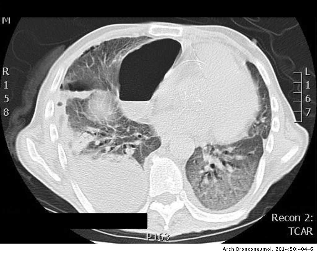
Right Lung Cavity With Bilateral Pleural Effusions Sgim Org from www.sgim.org Get expert advice on vaccines, medicines and more at docprime.com. Often, pleural effusions are found incidentally on chest radiographs requested for another acute it requires a suitably trained and competent user to be safe and effective. Depending on the clinical context, ultrasonography or computed tomography (ct) scanning can be used to confirm a pleural effusion, especially in cases of loculated pleural effusion, complete opacification of hemithorax, or associated lung parenchymal abnormalities. Ct scan of the chest of a patient with large loculated pleural effusion in his left thoracic cavity. More than one half of these massive pleural effusions are caused by malignancy; Clinical manifestations include chest pain, cough, and dyspnea. Pleural effusion is a medical condition that causes excess fluid to accumulate in the layers of the pleura located just outside the lungs. Ct scan of the chest.
A procedure that makes a series of detailed pictures of areas inside the body, taken from different angles.
Investigation of a unilateral pleural effusion in adults: Malignant pleural deposits or strange or atypical configurations of pleural fluid can be due to either adhesions (i.e. Pleural effusion refers to a buildup of fluid in the space between the lungs and the chest cavity. Pleural infection pleural inflammation pleural malignancy pleural fluid analysis findings: Improved after thoracentesis and diuresis. Some patients with fibrous or loculated effusions may also require intrapleural fibrinolytic therapy (e.g. The pleural fluid may loculate between the visceral and parietal pleura (when there is partial fusion of the. Ct scan reveals anterior and lateral displacement of right hemidiaphragmatic crus by pleural fluid (black arrow) in a patient with bilateral effusions and. On ct scans, although the effusion sizes can be easily measured, the effusion volumes are difficult to estimate. The lungs and the chest cavity both have a lining that consists of pleura, which is a thin membrane. More than one half of these massive pleural effusions are caused by malignancy; Positron emission tomography (pet) scan can help rule out extrathoracic disease that would preclude surgical. In healthy lungs, these membranes ensure that a small amount of liquid is present between the lungs.
Most likely secondary to left ventricular diastolic dysfunction loculated pleural effusion. A definite diagnosis of loculated pleural effusion is best established by ultrasonography or ct.


0 Komentar